Treatment of tricuspid valve regurgitation
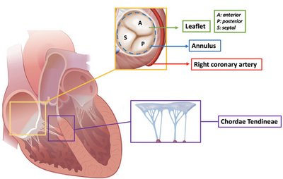
Tricuspid Valve
The tricuspid valve is located between the right atrium and right ventricle of your heart. In a normally functioning tricuspid valve, blood flows in a single direction from the right atrium into the right ventricle.
Tricuspid valve stenosis
Doctors refer to a narrowing of the tricuspid valve opening as tricuspid valve stenosis. If the narrowing causes blood to back up into the circulatory system, typical symptoms such as water retention (oedema), loss of appetite, nausea, vomiting and belching occur. Tricuspid valve stenosis is usually caused by rheumatic fever. As rheumatic fever hardly occurs in countries such as the UK and Ireland nowadays, tricuspid valve stenosis is also a rather rare disease. Other possible causes of tricuspid valve stenosis can be a tumor or a blood clot, for example.
Tricuspid valve regurgitation
Leakage of the tricuspid valve, also known as tricuspid regurgitation (TR), is a common condition. However, often only a small amount of blood flows back into the right atrium, so that no symptoms occur. Tricuspid regurgitation does not usually occur on its own, but as a result of another heart condition, such as heart failure (cardiac insufficiency), because when the right side of the heart enlarges, the tricuspid valve is pulled apart and can then no longer close properly. In severe cases, the heart can then no longer pump the blood effectively into the pulmonary circulation, causing it to back up into the systemic circulation.
Symptom Check
Symptoms
The signs and symptoms of tricuspid regurgitation depend on how advanced the insufficiency is and how quickly it has developed. Sometimes it causes few or no symptoms. However, if symptoms are present - especially in severe tricuspid regurgitation - they may include the following:
Shortness of breath, especially after exertion or when lying down
Drowsiness, brief unconsciousness, "blackness" before the eyes
Excessive water retention, swollen feet and ankles
Feeling of weakness and exhaustion, especially with increased activity (e.g. climbing stairs)
Irritable cough, which often worsens when lying down
Excessive urination at night
Loss of appetite
Palpitations, feeling of a fast, fluttering heartbeat

A leaky heart valve is tolerated by the body for a long time without those affected necessarily noticing. However, if the heart can no longer compensate for the backflowing blood by increasing its output, it backs up into the pulmonary circulation. When symptoms occur, there is often already advanced heart valve disease.
In advanced stages, tricuspid regurgitation can lead to the liver and kidneys no longer functioning properly and, if left untreated, can even lead to heart failure. Therefore, severe tricuspid regurgitation is a serious condition that requires timely treatment.
Diagnosis
If tricuspid regurgitation (TR) is suspected, various procedures can be used by your doctor to determine this specifically.

Auscultation (listening)
Listening to your heart with a stethoscope is the first thing to do if you suspect tricuspid regurgitation. The backflow of blood from the right ventricle through the diseased tricuspid valve into the right atrium of the heart can be heard clearly through the stethoscope.
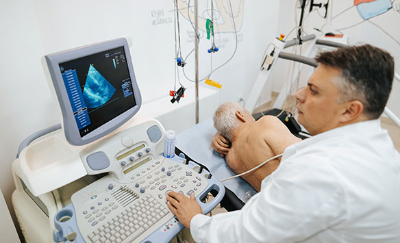
Echocardiography (echo)
One of the most important and informative examinations for detecting tricuspid regurgitation is an ultrasound examination of the heart, an echocardiogram. The doctor can see the blood flowing back from the right ventricle into the right atrium and the enlargement of the right atrium.
Echocardiography can be performed from the outside (transthoracic echocardiography) and also from the inside via the oesophagus (transoesophageal echocardiography). In a so-called stress echocardiography, the patient is exposed to a light load on the bicycle ergometer in order to accelerate the heartbeat, as it is difficult to assess the severity of the disease during an examination at rest. If this is not possible for health reasons, squeezing a foam ball several times quickly with the hand can also help to accelerate the heartbeat slightly and thus better visualise the tricuspid regurgitation.
Electrocardiogram examination (ECG)
ECG shows the electrical activity of the heart and helps to diagnose abnormal heart rhythm. This test is not specific for the tricuspid regurgitation diagnosis, but it is used to identify the functional state of your heart.
Find a heart centre
near you with
the clinic search
Search for a clinic
or cardiologist
near you.
Tricuspid Valve disease treatment
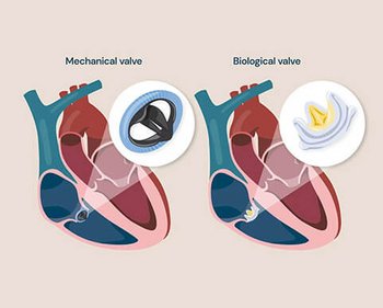
Treatment for tricuspid valve disease depends on the severity and type of the condition. Mild cases may be managed with medications such as diuretics to reduce fluid buildup and anticoagulants to prevent clots. In more advanced cases, surgical options are considered. These include valve repair, which is preferred when possible, or valve replacement using either mechanical or biological valves. Minimally invasive procedures, such as catheter-based techniques, may be suitable for high-risk patients. Treatment decisions are based on individual symptoms, overall health, and the presence of other heart conditions. Regular monitoring and specialist input are essential for optimal care.
PATIENT STORY - Lindel

How does TriClip™ therapy work?
Tricuspid TEER is a minimally invasive procedure that may be an option for patients who are too sick for surgery (also referred to as being high-risk for surgery). Unlike surgery, TEER does not require opening the chest and temporarily stopping the heart.
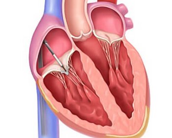
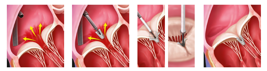
In the TEER procedure, the implant is placed on two or more leaflets of your tricuspid valve. This reduces tricuspid regurgitation, and the valve continues to open and close on either side of the implant, allowing blood to flow through. Sometimes, more than one implant is used on your tricuspid valve leaflets.
How does the procedure work?
The procedure does not require open heart surgery, thereby permitting real-time evaluation of the impact of TriClip™ Implant on TR and real-time positioning and repositioning to optimise TR reduction.
TriClip™ Implant/s is implanted with sterile techniques using fluoroscopy and echocardiography imaging. The TriClip System is carefully advanced under fluoroscopic guidance through the femoral vein to the heart, where it is positioned in the tricuspid valve. After the physician is satisfied with the TR reduction and tricuspid valve function, the implant is deployed, and the delivery system is removed from the patient's body.
The procedure is performed under a general anaesthetic.
Procedural steps
After the procedure
After percutaneous tricuspid valve repair with the TriClip™, the treating physicians will adjust the length of stay according to individual needs and health requirements.1

Most patients experience an improvement in their clinical symptoms (improvement in breathlessness and exercise tolerance) immediately after the procedure. Nevertheless, medication must still be taken after percutaneous tricuspid valve reconstruction, particularly to thin the blood and, if necessary, to treat any existing heart failure. It is very important that the doctor's instructions regarding medication that must be taken afterwards are followed in order not to jeopardise your health.1
After discharge from hospital, strenuous activities (such as lifting and carrying) should be avoided for at least 30 days. Most patients who have received a TriClip™ do not need any special help at home beyond the current care required for all other conditions.1
The attending physician will therefore give you the patient ID card after the procedure, which you should then always carry with you.
Videos with patient reports


Patient Story - Javier

Patient Story - Narcisa

Patient Story - Cruz
References
- Paul Sorajja et al. Transcatheter Repair for Patients with Tricuspid Regurgitation. N Engl J Med. 2023, DOI: 10.1056/NEJMoa2300525
- www.nejm.org/doi/pdf/10.1056/NEJMoa2300525
- patient.info/doctor/tricuspid-valve-disease
- www.nhs.uk/tests-and-treatments/heart-valve-replacement/why-its-done/
- www.bhf.org.uk/informationsupport/heart-matters-magazine/medical/treatments-for-heart-valve-disease
- guysandstthomasspecialistcare.co.uk/treatments/mitral-valve-repair-and-replacement/
9-UK-5-16496-01 09-2025 REV A