What is an echocardiogram?
An echocardiography, often referred to as a "heart echo", is an ultrasound examination (sonography) of the heart. A transducer emits waves at a high frequency, which then make various structures visible. These waves are invisible and do not cause any pain.
Echocardiography can be used to examine the heart valves and walls, as well as the pericardium. The pericardium is a protective sheath of connective tissue and surrounds almost the entire heart. Covered by a film of fluid, the heart can move in it with every beat, just like in a well-fitting glove. However, if the pericardium is filled with too much fluid or is hardened (e.g. due to calcification), this restricts the heart. These changes can be seen on a cardiac ultrasound.
If there are leaks (valve insufficiencies) or narrowing (valve stenosis), the heart is greatly enlarged or the walls are thickened, this can also be seen on ultrasound. Echocardiography is the method of choice for diagnosing mitral regurgitation or tricuspid regurgitation.
What procedures are available?
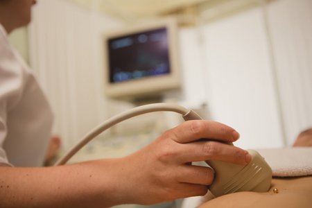
There are basically two different echocardiography methods: In transthoracic echocardiography (TTE), the transducer is placed on the chest (thorax) while you lie relaxed on your side. As bones strongly deflect the sound, the doctor always places the ultrasound probe between two ribs. A cool ultrasound gel is applied to the corresponding area beforehand, which improves the image quality.
If this examination does not reveal everything necessary, transoesophageal echocardiography (TOE) can be used as an alternative or supplement. "Trans" means "through", and the oesophagus is the gullet. As with a gastroscopy, a probe is inserted into the oesophagus with an ultrasound probe mounted on the tip. For this reason, this type of ultrasound examination is also known as a swallow echo. But don't worry: before the examination, the wall of the throat is locally anaesthetized with a spray - and if you wish, the doctor can also give you a sedative. The advantage of this method is that the oesophagus passes very close to the heart and you can see a lot more than with the transthoracic method.
Doppler ultrasound
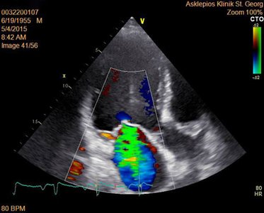
A Doppler ultrasound is a specialised mode of echocardiography that displays the velocity and direction of blood flow through the heart and blood vessels. It can even display blood flow in colour, making it easier to see how well the heart is working. This test helps doctors spot problems with the heart valves, such as leaks or narrowing. The test is safe, painless, and provides important information to help guide treatment and monitor heart health.
Ultrasound with contrast agent
Contrast-enhanced echocardiography is a special heart scan that helps doctors see the heart and its blood flow more clearly. A safe contrast agent, made of tiny bubbles, is injected into a vein. These bubbles improve the ultrasound images, making it easier to check how well blood is reaching the heart muscle and how the heart walls are moving. This test is especially helpful for spotting blocked arteries, checking heart muscle health, and guiding treatment decisions. It’s a safe and effective way to get a clearer picture of how your heart is working.
Resting and stress echocardiography
Echocardiography is usually first performed at rest. To see whether the blood flow in the coronary arteries or through the heart valves functions normally or is restricted under stress, the doctor can then perform a so-called "stress echo". The stress can either be performed directly on a bicycle ergometer or you can be given a drug that imitates stress. Exercise on a bicycle ergometer (or treadmill) is preferred, however, as the medication administered can alleviate the symptoms of heart disease and thus distort the results of the examination.
People who suffer from a leaky mitral valve (mitral regurgitation) or tricuspid valve (tricuspid regurgitation) do not necessarily have visible symptoms at rest. A stress echo can provide information here, as the symptoms then intensify.
Preparation for the examination
Patients do not need to take any special precautions before the TTE examination. However, if a TOE is planned, you must refrain from eating six hours beforehand and must not eat anything immediately afterwards.
If you are taking heart medication, you may not need to take it on the day of the examination. However, the doctor will inform you of this in good time during a consultation.
Risks of the individual procedures
The TTE examination is not dangerous as it does not interfere with the body's functioning; it can therefore also be carried out at any time in patients with circulatory problems, for example. TOE, on the other hand, can cause dizziness, nausea or weakness.
Some people also do not tolerate the contrast agent very well and may experience allergic reactions such as reddening of the skin and slowing of the heartbeat and/or a drop in blood pressure. However, these can usually be treated very well with medication.
Conclusion
Ultrasound examination of the heart is a very important tool for the reliable detection of heart diseases such as heart failure and valve defects. This procedure can accurately determine the type and extent of the impairment of the heart, which in turn is important for ensuring that those affected receive the right medication and treatment.
- References
- Magne J, Lancellotti P, Piérard LA. Exercise-induced changes in degenerative mitral regurgitation. J Am Coll Cardiol. 2010; 56(4):300-309. doi.org/10.1016/j.jacc.2009.12.073
- Magne J, Lancellotti P, Piérard LA. Exercise pulmonary hypertension in asymptomatic degenerative mitral regurgitation. Circulation. 2010; 122(1):33-41. doi.org/10.1161/CIRCULATIONAHA.110.938241
- Vohra, H.A., Burton, S., Yadav, R., Sayeed, R., Moorjani, N. Society of Cardiothoracic Surgery in Great Britain and Ireland guidance for adult mitral valve disease and interventions. BMJ Surgery, Interventions, & Health Technologies, 2025; 7:e000328. DOI: 10.1136/bmjsit-2024-000328
9-UK-5-16527-01 08-2025